Cell pilot results available
Three fields, numbered candidate masks, provisional round/flat labels and local WGA measurements. Attachment remains unverified; these are not validated cell counts.
Segmentation examples are awaiting review.Review image quality first, then validate cell detection and WGA classification. Candidate segmentation is available for pilot fields; validated attached-cell counts remain pending. No zero-valued biological results are implied.
Three fields, numbered candidate masks, provisional round/flat labels and local WGA measurements. Attachment remains unverified; these are not validated cell counts.
Segmentation examples are awaiting review.Priority audit: distinguish suspected focus loss from true empty fields. Camera dust alone cannot establish sample focus. Review acquisition chronology and original z planes; exclude failed-focus fields from counts and from confirmed empty controls.
Read the focus-first pilot planCompare median, minimum, maximum and axial brightfield evidence
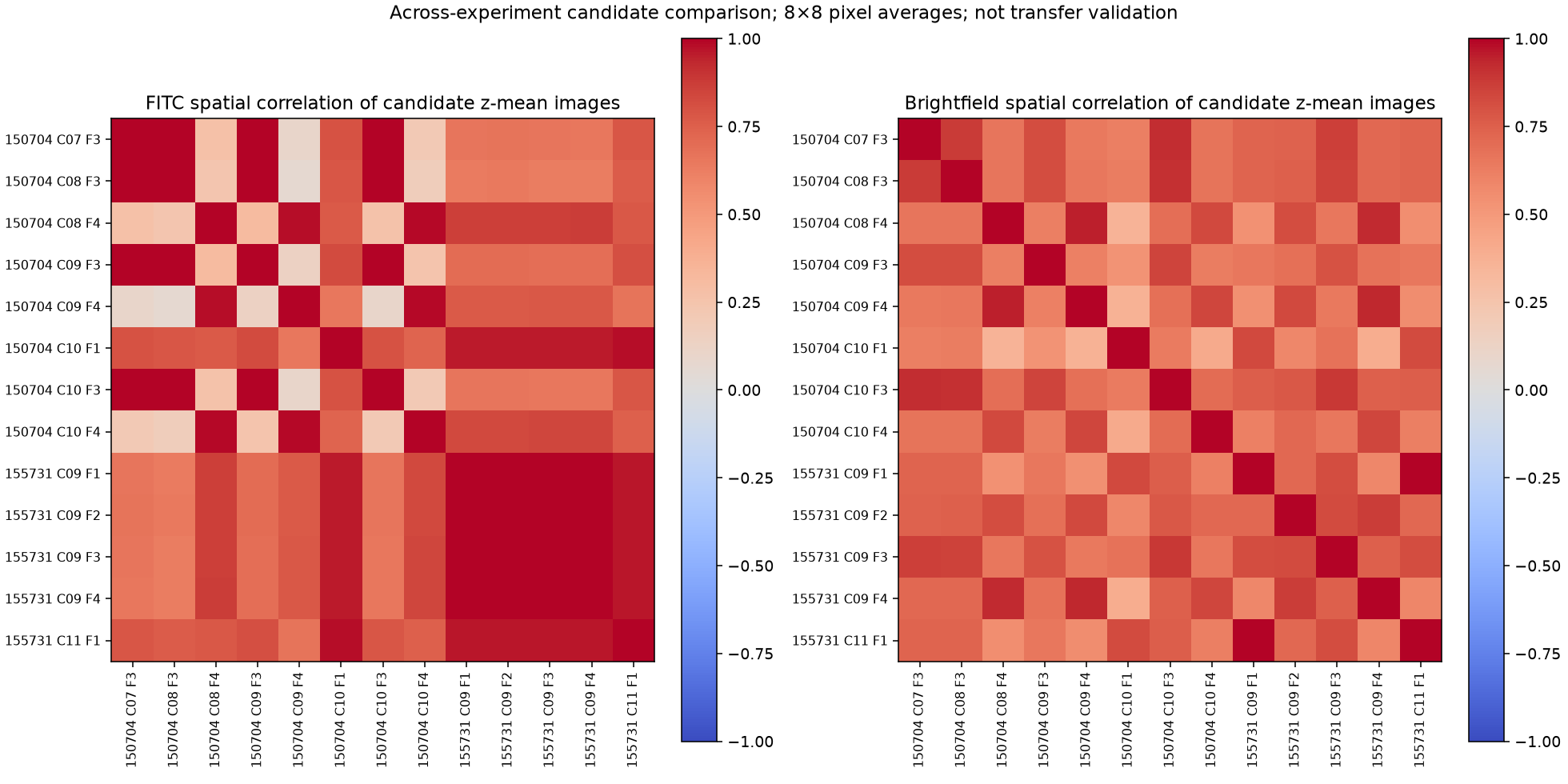
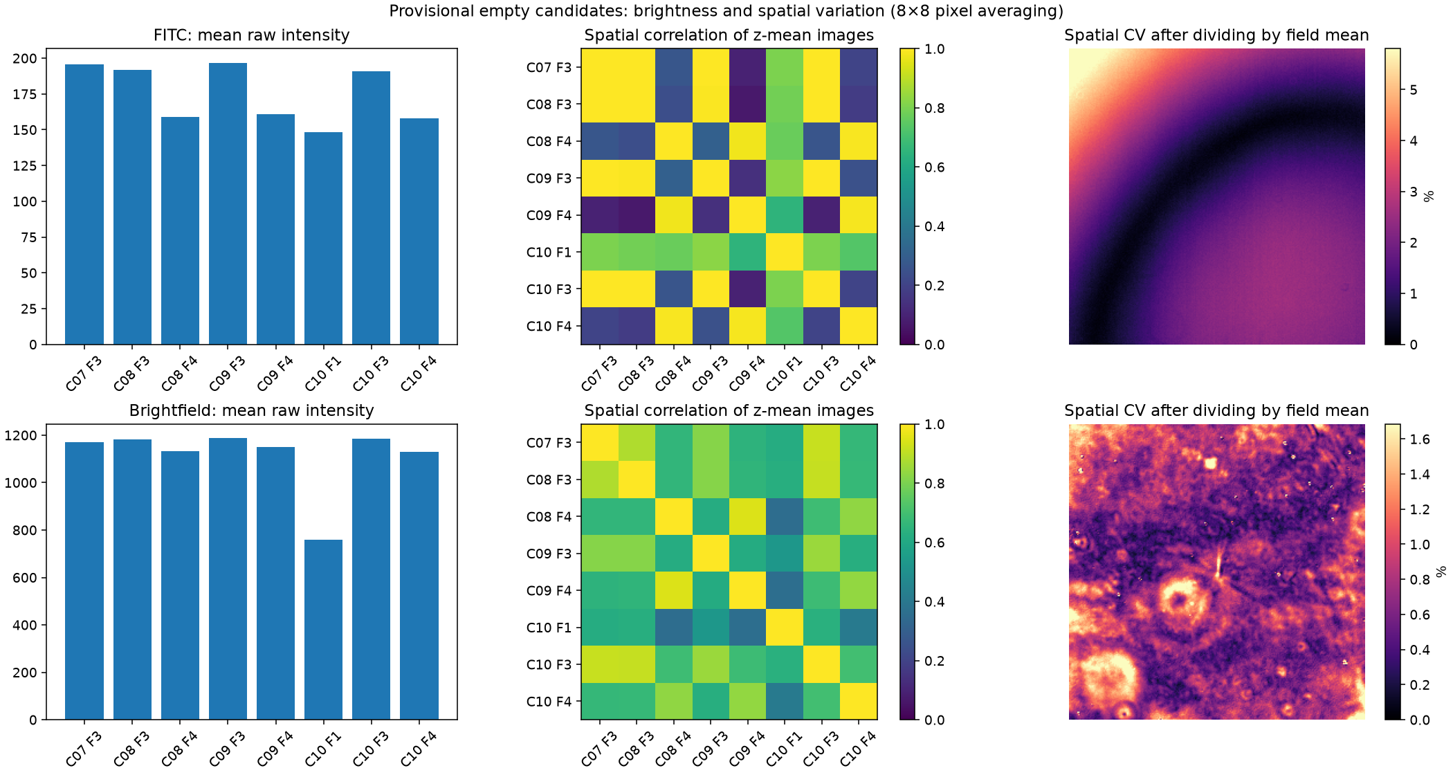
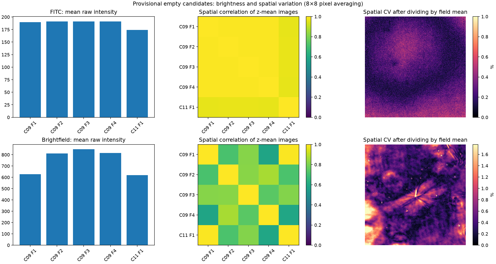
These are descriptive comparisons of provisional background-only fields. Empty versus missed focus remains unresolved, so they are not yet validated calibration controls.
Review brightfield correction and cell-outline crops
Review fluorescence background subtraction: before / after pilot
Segmentation examples are awaiting review.
Next: numbered examples to agree on round/long morphology, attachment evidence, detectable WGA and ambiguous cases. Shape measurements, cell labels and well-level counts will appear here after validation.
Pending reviewed examples. Distinguish cluster membership and central versus broad fluorescence without double-counting cells.
Notebook refreshed 2026-10-24T00:43:07-04:00 (Toronto). Measurements describe the saved snapshot; refresh after copying more data.